
“See-through” larvae of zebrafish reveal how wound healing leads to skin cancer.
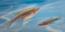
Genetically modified larvae of zebrafish were studied by the researchers in the United Kingdom and Denmark to watch the relationship between wound-associated inflammation and melanoma.
The cellular events and changes were observed by live imaging with a special confocal laser-scanning microscope.
The experiments showed that neutrophils, the protective inflammatory cells of the body’s immune system, gets diverted from an induced wound to any nearby precancerous skin cells.
They were able to locate that a specific type of signaling molecule released by neutrophils named prostaglandin E2 is part of the signal that drives the splurge of cell growth linked to the cancer.
The newly arrived neutrophils cause rapid division of these skin cells which may cause them to progress to melanoma.
Scientists have known for some time that inflammation is one of the ten hallmarks of cancer which is also known as the “wound that does not heal.” High levels of neutrophils were also detected in human clinical samples of melanomas that had been obtained from individuals whose cancers had open ulcers.
It was concluded that neutrophils was linked to increased proliferation of melanoma cells and poor survival which suggests that these findings in fish may have considerable relevance to cancer patients. These experiments suggest that several strategies might improve outcomes for patients including the possible use of therapeutics to dampen damage-induced inflammatory responses.
Minimally invasive surgery is beneficial to cancer patients in many situations and often the preferred treatment but there are cases where all cancerous tissue cannot be removed and therefore the inflammatory response might influence the remaining cancer cells in the body.
Further work is in progress to better understand the relationship between the inflammatory response and skin cancer in the zebrafish model system as the findings of the study may have implications for cancer surgery.
For more information please visit:
Comments are closed.