
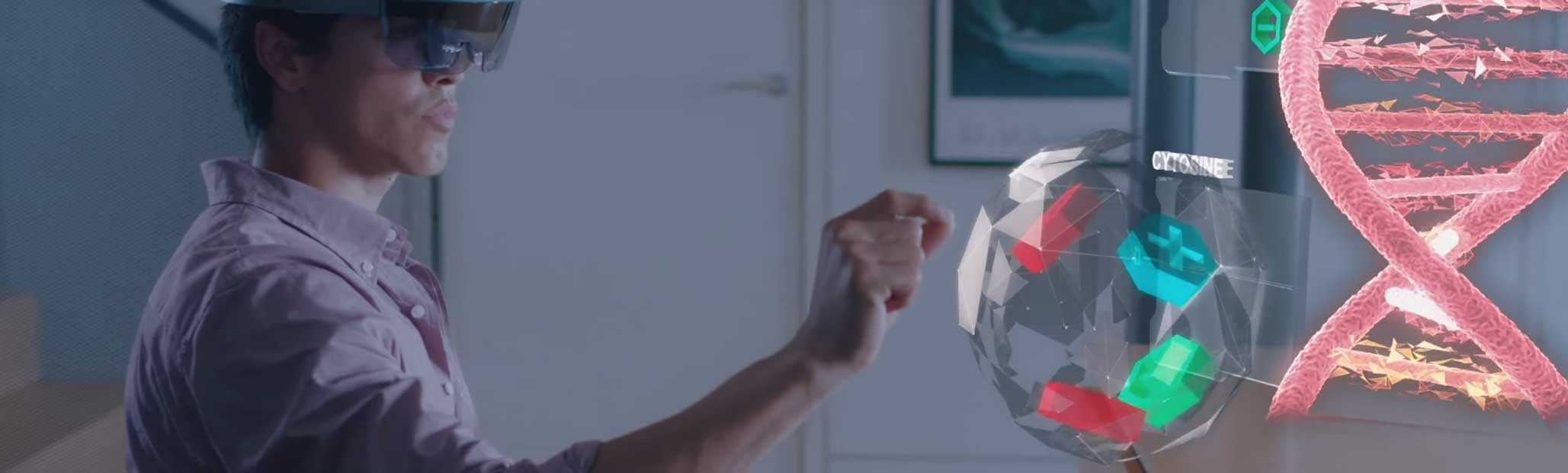
Cleveland Clinic has partnered with Case Western Reserve University to release a Microsoft HoloLens app that allows users to explore the human body using augmented reality technology. The HoloLens is a headset that superimposes computer generated 3D graphics onto a person’s field of view, essentially blending reality with virtual reality.
The HoloAnatomy app lets people explore a virtual human, to walk around it looking at details of different systems of the body, and to select which are showing. Even some sounds are replicated, such as that of the beating heart.
Case Western Reserve and Cleveland Clinic broke ground on a 485,000-square-foot Health Education Campus, a space designed to support interprofessional learning and offer the most advanced technology available.
As part of creating what Davis called a “state-of-the-future” building, the new space will not include any of the traditional cadaver-filled laboratories that for decades have housed anatomy classes. Instead, just as Mlakar and Eastman did Wednesday, medical students will don HoloLens headsets to see the body’s organs and systems.
The pair began with a hologram of the digestive system, with labels on organs including the stomach, gall bladder and liver. Davis asked them about the pancreas, which, nestled behind the stomach, cannot be seen from the front of the body.
Thankfully with HoloLens, Mlakar said, making a small hand gesture that caused the holographic body to rotate 180 degrees to reveal the organ, “it’s really easy to get the best view of things.”
It’s also easy to engage with an instructor hundreds of miles away, as Professor Griswold demonstrated when a simple 3-D head and arm representing him entered the sold-out auditorium.
“This is our new system,” he said, “which allows me to teach and interact with you, even though I’m not there. … This is really changing what it means to be ‘in class.’”
Based on an actual patient MRI from Case Western Reserve Professor Cameron McIntyre’s lab, Griswold guided Eastman and Mlakar through an examination of the white-matter tracts of the brain, fibers that allows messages to travel from one area to another. Based on an actual patient’s MRI, the hologram color-coded the fibers by direction, and included a bright red mass that Griswold identified as a tumor.
Credit : Cleveland Clinic and Case Western Reserve University