The cerebrospinal fluid from young mice is awash with factors that keep the brain sharp. Now, scientists led by Tony Wyss-Coray at Stanford University in Palo Alto, California, report that when injected into the brains of old mice, CSF from young mice, or young people, revved expression of a host of oligodendrocyte genes within the hippocampus, counteracting the slump in proliferation and function of the myelin-making cells that typically occurs with age.
The researchers tied the effects to fibroblast growth factor 17 in the young CSF, which increased expression of serum response factor in oligodendrocytes, enhancing their proliferation and myelin production.
“This work demonstrates the rejuvenating capacity of young CSF, and highlights oligodendrocyte lineage cells as a potential target for therapeutic strategies to prevent age-associated cognitive decline,” wrote Hansruedi Mathys of the University of Pittsburgh.
The findings are the latest in a long line of studies from Wyss-Coray’s lab that identified factors in young animals that influence aging of the brain (for review, Pluvinage and Wyss-Coray, 2020). Previously, the group reported that human umbilical cord blood, and plasma from people in their 20s or from 3-month-old mice, boosts neurogenesis, neuronal plasticity in the hippocampus, and memory in aged mice (May 2014 news; Apr 2017 news; Sep 2019 news).
Compared to plasma, cerebrospinal fluid is far more intimately connected to brain. Churned out in the choroid plexus and imbued with neuroprotective factors, CSF works its way through the brain via the glymphatic system (Aug 2012 news). Its composition also changes with age. Might CSF components from the young rejuvenate the aging brain? First author Tal Iram and colleagues collected CSF and infused it directly into the right lateral ventricles of 20-month-old animals daily for a week. They started the infusions just after they gave the animals a foot shock and tested their memories three weeks after the shock. Compared to old mice infused with artificial CSF, those infused with the fluid from 3-month-old mice were more adept at remembering where they had previously received series of unpleasant foot shocks three weeks prior—a stretch of time considered “remote” for a mouse. This suggested that the young CSF improved the animals’ remote recall, a type of memory that depends on the hippocampus. Given the timing of the infusion, the researchers believe the CSF helped the mice consolidate memories.
To investigate this improvement, the researchers looked for changes in the hippocampal transcriptome using RNA-Seq. They found a rise in transcripts encoding oligodendrocyte genes, including those that drive differentiation of the myelin-producing cells from oligodendrocyte precursors (OPCs), as well as an uptick in expression of major components of myelin itself. Neither artificial CSF, nor CSF from aged mice, triggered these responses. Furthermore, the percentage of actively proliferating OPCs in the hippocampus more than doubled when mice were infused with young CSF. Notably, human CSF pooled from donors in their 20s triggered a similar OPC proliferative response, while CSF pooled from donors in their 70s only coaxed half as many cells to divide. Curiously, young CSF did not spur OPC proliferation in the cortex, suggesting that hippocampal OPCs were particularly responsive.
Did these proliferating OPCs mature into bona fide myelin-makers? Indeed, the researchers found that three weeks after infusion with young CSF, levels of myelin basic protein, as well as numbers of myelinated axons, had increased in the molecular layer of the hippocampus.
How did young CSF promote an oligodendrocyte renaissance in aged mice? A clue came from the first batch of transcripts to rise in response to CSF. Using metabolic labeling to tag freshly made transcripts in cultured OPCs, Iram found that within one hour of treatment with young CSF, expression of serum response factor (SRF) was more strongly upregulated than any other gene. Expressed in multiple organs throughout the body, SRF binds to serum response element (SRE) promoter sequences, activating genes that help coordinate cytoskeletal arrangements involved in all manner of cellular functions, including motility and proliferation. In neurons, SRF orchestrates the growth of axons and aids in synapse formation. What role does the transcription factor play in oligodendrocytes? Iram said they are looking into that, but for the purposes of this study, the researchers found that SRF was necessary for OPCs to proliferate in response to young CSF. Moreover, expression of SRF in mouse hippocampal OPCs waned markedly as the animals aged. Digging through data from published human studies, the authors found that SRF expression also tapers in the brains of people with AD (Mathys et al., 2019; Zhou et al., 2020).

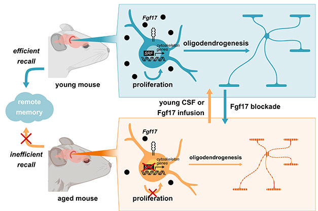
Young vs. Old. Abundant Fgf17 (black dots) in the CSF of a young mouse (top), promotes SRF-driven gene expression, enhancing oligodendrogenesis, myelination of axons, and sharp memory. In old mice, Fgf17 wanes, gene expression and myelin production fall, and memory falters (bottom). Infusions of young CSF or Fgf17 rejuvenates old mice (orange arrow), while blocking Fgf17 in young mice makes them behave like old (blue arrow). [Courtesy of Iram et al., Nature, 2022.]
What factors within young CSF dialed up SRF expression? The authors speculated that hundreds of proteins within CSF could do the trick. In fact, many SRF target genes also feedback to induce SRF itself, and the researchers leveraged this to whittle down the list of potential suspects. By cross-referencing CSF proteomics datasets with lists of known SRF target genes, they came up with 35 potentials. Among them, Iram found that two—Fgf8 and Fgf17—most strongly induced SRF expression in human kidney cells. Because it is preferentially expressed in the brain and reportedly drops in human CSF with age, the researchers investigated Fgf17 further. They found it expressed in neurons in the mouse cortex and hippocampus, and that this expression drops dramatically with age. However, they could not detect the growth factor in their mouse CSF samples. Still, when infused into aged mice, Fgf17 enhanced SRF expression in hippocampal OPCs, ramped up OPC proliferation, and even improved memory, just like the young CSF. Infusion of Fgf17 antibodies into young mice worsened the animals’ performance on memory tests, suggesting the factor plays a broad role in memory formation. Notably, blocking the growth factor in OPC cultures stymied their proliferation.
In all, the findings suggest that Fgf17 and potentially other growth factors within young CSF promote critical functions of oligodendrocytes, and that loss of these factors with age may lead to the erosion of oligodendrocyte function and myelination. While myelin in the brain is mostly wrapped around axons early in life, recent studies suggest that fresh production of this fatty sheath, spurred by neuronal activity, is important for learning and memory throughout life. Iram noted that myelin production increases into adulthood before starting to wane as people enter their 60s. Together, these studies implicate flagging oligodendrocyte function in age-related cognitive decline, she said.
To Ragnhildur Thura Káradóttir of the University of Cambridge, U.K., the findings add to mounting evidence that oligodendrocytes and myelin maintenance play a prominent role in age-related memory loss and neurological disease. In the past, the role of oligodendrocytes in these processes has been underappreciated, she noted. Iram’s study goes a step further, by showing that the brain’s environment—in this case, factors in the CSF—may be capable of driving, or slowing, these age-related changes, she told Alzforum. Káradóttir was intrigued that Fgf17 enhanced oligodendrocyte function, but thinks it likely only one piece of a much larger picture of CSF/oligodendrocyte relations.
Vivek Swarup of the University of California, Irvine, agreed that future work will be needed to paint a more comprehensive picture of how factors in CSF influence oligodendrocyte development and function. He wondered how the findings translate to what happens in Alzheimer’s and other neurodegenerative diseases. “Will young CSF also curb memory loss caused by neurodegenerative disease?” he asked. The answer to that question could be exciting from a therapeutic standpoint, he said.
Li-Huei Tsai and Leyla Akay of Massachusetts Institute of Technology agreed (comment below). “Understanding how SRF and Fgf17 promote myelination, and determining whether this can be leveraged to treat neurodegenerative diseases associated with demyelination and white-matter injury, will make for exciting future research,” they wrote.
In a Nature News & Views, Miriam Zawadzki and Maria Lehtinen of Boston Children’s Hospital noted that the paper raises a provocative hypothesis about protein and fluid distribution throughout the brain. “Unexpectedly, Iram and colleagues found that FGF17 in the CSF isn’t sourced by the choroid plexus, but, instead, by youthful neurons themselves, providing evidence that neuron-based signals are delivered by the CSF,” they wrote. “How FGF17 is distributed in the CSF and delivered to target cells in the hippocampus presents a new direction of research.”—Jessica Shugart
Credit : University of California, Irvine
Credit : Stanford University in Palo Alto, California
Credit : https://www.alzforum.org/
