
Researchers at the University of Calgary have made a groundbreaking observation confirming the clear existence of biophotons—ultra-weak photon emissions produced by biological systems—in two plant species and live mice. This revelation not only substantiates the long-speculated phenomenon of biological light but also opens new frontiers in understanding cell communication, oxidative stress, and potentially even consciousness. The findings suggest that all living organisms, including humans, might emit a constant, invisible glow—one that lasts until the very moment of death.
Biological Significance of Biophotons
The implications of biophoton detection are vast and multifaceted:
A. Cellular Communication
- Evidence supports that biophotons might play a role in cell-to-cell signaling, possibly acting as a non-chemical method of long-range communication.
- The coherence of biophoton emissions hints at laser-like properties in biological systems.
B. Oxidative Stress Biomarker
- Emissions are closely correlated with reactive oxygen species (ROS) levels, making biophotons a promising, non-invasive biomarker for:
- Cancer detection
- Aging studies
- Neurodegenerative diseases
- Immune response evaluation
C. Consciousness and Brain Activity
- Biophoton emissions from the brain—especially from neurons and microtubules—have been proposed as a possible mechanism for neural synchronization or even quantum brain theories.
The phenomenon of biological ultraweak photon emission (UPE), that is, extremely low-intensity emission (10–103 photons cm–2 s–1) in the spectral range of 200–1000 nm, has been observed in all living systems that have been examined. Here, we report experiments that exemplify the ability of novel imaging systems to detect variations in UPE for a set of physiologically important scenarios.
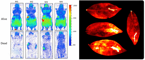
We use electron-multiplying charge-coupled device (EMCCD) and charge-coupled device (CCD) cameras to capture single visible-wavelength photons with low noise and quantum efficiencies higher than 90%. Our investigation reveals significant contrast between the UPE from live vs dead mice. In plants, we observed that an increase in the temperature and injuries both caused an increase in UPE intensity.
Imaging Ultraweak Photon Emission from Living and Dead Mice and from Plants under Stress
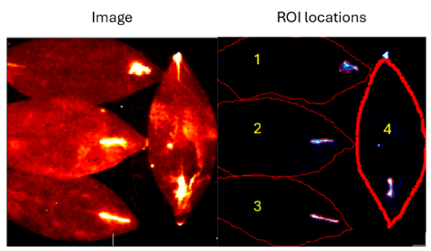
Moreover, chemical treatments modified the UPE emission characteristics of plants, particularly the application of a local anesthetic (benzocaine) to injury, which showed the highest emission among the compounds tested. As a result, UPE imaging provides the possibility of non-invasive label-free imaging of vitality in animals and the responses of plants to stress.
Biophotons are extremely low-intensity light particles (photons) emitted from the cells of living organisms. Unlike bioluminescence, which is visible to the naked eye (e.g., fireflies), biophotons are about 1,000 times weaker than the sensitivity threshold of the human eye, usually in the range of 200 to 800 nm.
Imaging Technology and Methods
The University of Calgary team utilized a cutting-edge time-resolved photon detection system integrated with custom-developed software for real-time visualization. The core imaging protocol included:
- Dark adaptation of subjects before imaging to eliminate residual light
- Use of optical bandpass filters to isolate biologically relevant wavelengths
- Biological shielding to rule out thermal or chemiluminescent noise
First theorized in the early 20th century and later studied in depth by researchers like Fritz-Albert Popp in the 1970s, biophotons are now recognized as real phenomena. They are believed to be produced during metabolic processes, particularly through reactive oxygen species (ROS) and other radical-mediated oxidative reactions in cells.
University of Calgary Research Overview
In a 2024 publication from the University of Calgary’s Faculty of Science, researchers employed advanced photon counting instrumentation to conclusively detect biophoton emissions in Arabidopsis thaliana, Pisum sativum (common pea plant), and live laboratory mice. Their instrumentation setup included:
- High-sensitivity photomultiplier tubes (PMTs)
- Dark chamber setups for light isolation
- Spectral filters for emission wavelength analysis
- Cryogenically cooled CCD cameras for imaging
The study focused on quantifying and visualizing the emission spectrum, rate, and spatial distribution of biophotons from the surface of living tissues under stress and normal conditions.
Key Observations
1. Presence of Biophoton Emission in Plants
- Arabidopsis and pea plants both showed clear, spontaneous photon emission.
- Emission intensity increased significantly during periods of oxidative stress, such as during leaf injury or exposure to UV-A radiation.
- Wavelengths peaked in the green to near-infrared spectrum (~550–800 nm), consistent with mitochondrial and chloroplast oxidative reactions.
2. Biophoton Emission in Mice
- Live anesthetized mice were observed to emit low-level light, with the strongest emissions localized in head, chest, and abdominal areas—where metabolic activity is concentrated.
- Emission levels decreased rapidly post-mortem, with a distinct decay pattern indicating cessation of metabolic processes.
- This provides a visual, optical marker for the moment of death at the cellular level.
3. Lifespan and Photon Emission
- In both plants and animals, a gradual decline in biophoton emission was recorded as biological systems approached death.
- This supports the idea that “glowing until we expire” may be a literal truth—albeit invisible without scientific instrumentation.
The University of Calgary’s experimental validation of biophoton emissions in both flora and fauna marks a turning point in biophysics and systems biology. Their discovery not only affirms that living organisms emit a silent, continuous light but also raises profound questions about the role of this radiation in life’s processes. As research advances, we may find that life is not only about biochemistry—but also about bio-optics. And perhaps, just perhaps, every living thing truly glows until it dies.