3D Bioprinting is a form of additive manufacturing that uses cells and other biocompatible materials as “inks”, also known as bioinks, to print living structures layer-by-layer which mimic the behavior of natural living systems.
Three dimensional bioprinting is the utilization of 3D printing–like techniques to combine cells, growth factors, and biomaterials to fabricate biomedical parts that maximally imitate natural tissue characteristics.
3D bioprinting can be used to reconstruct tissue from various regions of the body. Patients with end-stage bladder disease can be treated by using engineered bladder tissues to rebuild the damaged organ. This technology can also potentially be applied to bone, skin, cartilage and muscle tissue.
scientists have printed mini organoids and microfluidics models of tissues, also known as organs on chips.
Researchers have been using 3D–printing techniques in hopes of developing tissues that can be transplanted into humans.
Allevi bioprinters – Allevi bioprinters enable users to experiment with any biomaterial, work with any cell line, and print in any geometry. Print anything from brain to bone with Allevi bioprinters.
The Allevi 2 is built around a compressed air pneumatic system, making it very easy to achieve clean starts and stops in printing. Pressure ranges between 1 and 120 PSI accommodate a wide range of viscous materials.
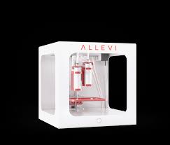
With temperature control from room temperature to 160° C, the Allevi 2 bioprinter is able to print a wide range of bioinks and cells including gelatin methacrylate, PCL, PLGA, Hyperelastic Bone, fibroblasts, and mesenchymal stem cells (hMSC)– just to name a few.
With the Allevi 2 bioprinter you can choose to cure biomaterials in either visible or ultraviolet light, using LAP or another photoinitiator. We will build the printer to your specification when you buy – 405nm crosslinking LEDs are standard, with 365nm as an alternate option.
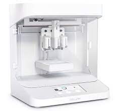
Cellink 3d bioprinter –
THREE DIMENSIONAL BIOPRINTING OF A VASCULARIZED AND PERFUSABLE SKIN GRAFT USING HUMAN KERATINOCYTES, FIBROBLASTS, PERICYTES, AND ENDOTHELIAL CELLS.

Multilayered skin substitutes comprising allogeneic cells have been tested for the treatment of nonhealing cutaneous ulcers. However, such nonnative skin grafts fail to permanently engraft because they lack dermal vascular networks important for integration with the host tissue. In this study, we describe the fabrication of an implantable multilayered vascularized bioengineered skin graft using 3D bioprinting.
The graft is formed using one bioink containing human foreskin dermal fibroblasts (FBs), human endothelial cells (ECs) derived from cord blood human endothelial colony-forming cells (HECFCs), and human placental pericytes (PCs) suspended in rat tail type I collagen to form a dermis followed by printing with a second bioink containing human foreskin keratinocytes (KCs) to form an epidermis.
In vitro, KCs replicate and mature to form a multilayered barrier, while the ECs and PCs self-assemble into interconnected microvascular networks. The PCs in the dermal bioink associate with EC-lined vascular structures and appear to improve KC maturation. When these 3D printed grafts are implanted on the dorsum of immunodeficient mice, the human EC-lined structures inosculate with mouse microvessels arising from the wound bed and become perfused within 4 weeks after implantation. The presence of PCs in the printed dermis enhances the invasion of the graft by host microvessels and the formation of an epidermal rete.