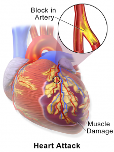
Researchers have discovered that a particular protein, Fstl1, plays a key role in regenerating dead heart-muscle cells.
Scientists at the Stanford University School of Medicine and their colleagues have enabled damaged heart tissue in animals to regenerate by delivering a protein to it via a bioengineered collagen patch.
There is currently no effective treatment to reverse the scarring in the heart after heart attacks. Heart attacks cause millions of deaths annually worldwide and are predicted to skyrocket in the next few decades tripling by 2030. In a heart attack, cardiac muscle cells, called cardiomyocytes, die from a lack of blood flow. Replacing those dead cells is vital for the organ to fully recover. Unfortunately, the adult mammalian heart does not regenerate effectively, causing scar tissue to form. Protein helps regenerate dead heart muscle.
About 735,000 Americans suffer a heart attack each year. Many victims now survive the initial injury, thanks to advances in early treatment, but the resulting loss of cardiomyocytes can lead to heart failure and possibly death. Consequently, most survivors face a long and progressive course of heart failure, with poor quality of life and very high medical costs. Various methods of transplanting healthy muscle cells into a damaged heart have been tried, but have yet to yield consistent success in promoting healing.
Previous heart regeneration studies in zebrafish have shown that the epicardium is one of the driving factors for healing a damaged heart. Reserchers wanted to know what in the epicardium stimulates the myocardium, the muscle of the heart, to regenerate.
Since adult mammalian hearts do not regenerate effectively, they also wanted to know whether epicardial substances might stimulate regeneration in mammalian hearts and restore function after a heart attack. They pinpointed Fstl1, a protein secreted by the epicardium, as a growth factor for cardiomyocytes. Not only did this protein kick-start the proliferation of cardiomyocytes in petri dishes, but the researchers were surprised to find that it was missing from damaged epicardial tissue following heart attacks in humans.
The researchers set out to reintroduce the protein back into the damaged epicardial tissue of mice and pigs that had suffered a heart attack. They did this by suturing a bioengineered patch, loaded with Fstl1, to the damaged tissue. The patches were made of natural material known as collagen that was structurally modified to mimic certain mechanical properties of the epicardium. Because the patches are made of acellular collagen, meaning they contain no cells, recipients do not need immunosuppressive drugs to avoid rejection. With time, the collagen material gets absorbed into the organ. The researchers believe that the elasticity of the material, which resembles that of the fetal heart, is key to providing a hospitable environment for muscle regrowth. New blood vessels regenerated there as well. Within two to four weeks of receiving the patch, heart muscle cells began to proliferate and the animals progressively recovered heart function The hope is that a similar procedure could eventually be used in human heart-attack patients who suffer severe heart damage.
But by placing a bioengineered patch that mimics membrane tissue and acts as a source of FSTL1 on damaged mouse and pig hearts, the study authors found heart muscle cells started growing and spreading, improving heart function and survival. Two weeks later the hearts began to grow fresh muscle cells and new blood vessels, while showing signs of pumping more effectively.
The results were remarkable. The protein not only reduced scarring, but it rebuilt the damaged heart as well. Furthermore, the patches contained no cells, but were specifically made of the main structural protein in the extracellular space named “collagen”. A group of researchers managed to embed a naturally-occurring protein into a patch which can help heal muscles damaged by heart attacks.
The heart rate of a pig which suffered from heart attack dropped from 50% to 30% in the left ventricle. They then designed a collagen-based material coated in FSTL1, and placed it on the hearts of pigs and mice forced to have a heart attack. This would change future treatment, and lead to the reduction of long-term damage in the event of a heart attack. These cells led to less scarring, and the animal’s hearts returned to near normal function very soon after the procedure.
The protein in question is called Follistatin-like 1 (FSTL1), and initial tests in lab environment showed that the protein can stimulate cultured heart muscle cells to divide. It is commercially viable, clinically attractive and you don’t need immunosuppressive drugs.
The new creation could be set to enter human clinical trials as early as 2017.
This finding opens the door to a completely revolutionary treatment.
For more information please visit: www.med.stanford.edu

