
The analysis of MRI scans of the ageing hearts of nearly 3,000 adults was led by investigators at Johns Hopkins.’ Our results are a striking demonstration of the concept that heart disease may have different pathophysiology in men and women and of the need for tailored treatments that address such important biologic differences,’ said João Lima of the Johns Hopkins University School of Medicine, who led the research. Published in the journal Radiology, this is believed to be the first long-term follow-up using MRI, showing how hearts change as they age.Previous studies have assessed heart changes over time using ultrasound, but, the researchers say, MRI scans tend to provide more detailed images and more reliable information about the structure and function of the heart muscle.
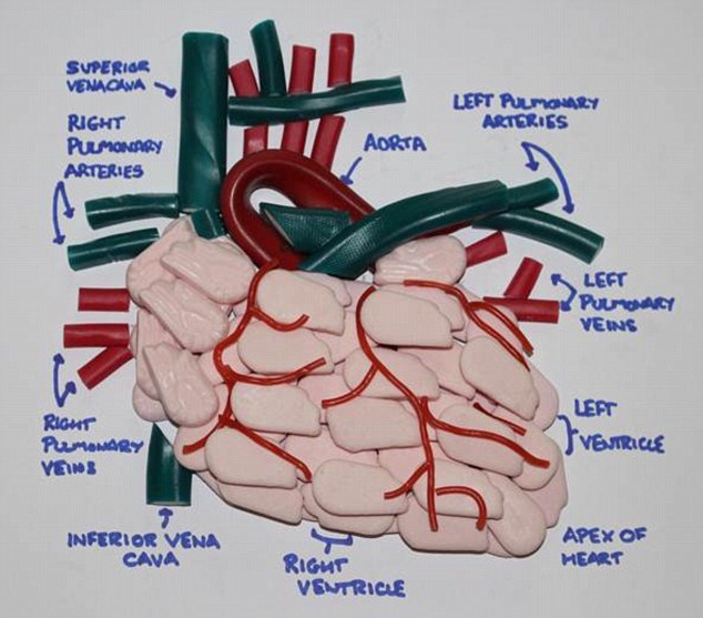
In both sexes, the main heart chamber, the left ventricle which fills with and then forces out blood gets smaller with time. As a result, less blood enters the heart and less gets pumped out to the rest of the body. But in men, the study reveals, the heart muscle that encircles the chamber grows bigger and thicker with age, while in women, it get retains its size or gets somewhat smaller.’Thicker heart muscle and smaller heart chamber volume both portend heightened risk of age-related heart failure but the gender variations we observed mean men and women may develop the disease for different reasons,’ says lead investigator John Eng, M.D., associate professor of radiological science at the Johns Hopkins University School of Medicine
A condition that affects more than five million Americans, heart failure is marked by the gradual ‘floppiness’ and weakening of the heart muscle and eventual loss of pumping ability. To lower the risk, cardiologists often prescribe medications designed to reduce the thickness of the heart muscle over time and boost cardiovascular performance. But the finding that in women, the heart muscle tends to shrink or remain the same size means they may not derive the same benefit from such treatments, researchers say.The research team cautions that its study was not designed to find what exactly fuels the differences in cardiac physiology between the sexes but says this ‘fascinating discrepancy’ demands further investigation to figure it out.
For the study, researchers analyzed MRI scans performed on nearly 3,000 older adults, ages 54 to 94, without preexisting heart disease. Participants were followed between 2002 and 2012, at six hospitals across the United States where each one of them underwent MRI testing at the beginning of the study and once more after a decade.The MRI scans provided researchers with 3-D images of the heart’s interior and exterior, allowing them to determine the size and volume of the heart muscle. Adding these to the already known density of the muscle, they were able to calculate its weight.
Over a period of 10 years, the weight of the heart’s main pumping chamber, the left ventricle, increased by an average of 8 grams in men and decreased by 1.6 grams in women. The heart’s filling capacity, which is marked by the amount of blood the left ventricle can holds between heartbeats, declined in both sexes but more in women, by about 13 milliliters, compared with just under 10 milliliters in men.The differences in size, volume and pumping ability occurred independently of other risk factors known to affect heart muscle size and performance, including body weight, blood pressure, cholesterol levels, exercise levels and smoking.
For more information please visit:: www.rsna.org
Comments are closed.