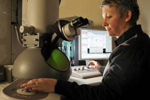
Deborah Kelly, a biologist at Virginia Tech Carilion Research Institute, has developed a “microchip-based toolkit” to watch the breast cancer affiliated BRCA1 gene act inside a human breast cancer cell. This new technology allows scientists to watch cancer cells in action at unprecedented resolution. They can now peer closely into the world of cells and molecules within a native, liquid environment. Kelly and colleagues have developed a way to isolate biological specimens in a flowing, liquid environment while enclosing those specimens in the high-vacuum system of a transmission electron microscope (TEM).
The TEM liquid-flow holder, developed by Protochips Inc. of Raleigh, N.C., accommodates biological samples between two semiconductor microchips that are tightly sealed together. These chips form a microfluidic device smaller than a Tic Tac. This device, positioned at the tip of an EM specimen holder, permits liquid flow in and out of the holder. When these chips are coated with a special affinity biofilm that Kelly developed, they have the ability to capture cells and molecules rapidly and with high specificity. This system allows researchers to watch at unprecedented resolution biological processes as they occur, such as the interaction of a molecule with a receptor on a cell that triggers normal development or cancer.
“With this new technology, we can capture and view the native architecture of cells and their surface protein receptors while learning about their dynamic interactions, such as what happens when cells interact with pathogens or drugs,” said Kelly. “We can now isolate cancer cells, for example, and view the early events of chemotherapy in action.” Kelly had previously worked with colleagues at Harvard Medical School to develop a way to capture protein machinery in a frozen environment. “But life moves,” said Kelly. “It’s better if biological processes don’t have to be paused or frozen in order to be studied, but can be viewed in dynamic and life-sustaining liquid environments.”
Kelly’s affinity capture device, in combination with high-resolution TEM, helps bridge the gap between cellular and molecular imaging, allowing researchers to achieve spatial resolution as high as two nanometers. “This device allows us to see new features on the surface of live cancer cells, providing new targets for drug therapy,” Kelly said. “With this resolution, scientists may even be able to visualize disease processes as they unfold.”
BRCA1 is one of the most important genes involved in the study of breast cancer. Ideally, BRCA1 acts as a sort of quality control specialist that oversees cell division. If the cells are damaged in this process, BRCA1 goes in and corrects the error by sending in special proteins. But if a patient has a mutation in the BRCA1 gene, her or his quality control system is weakened, and cell errors can become tumors. That’s why a BRCA1 mutation is strongly correlated with breast and ovarian cancers. Americans have recently become more aware of the BRCA mutations (BRCA1 and BRCA2) following Angelina Jolie’s announcement that she had tested positive for a BRCA mutation, and her elective mastectomy that followed. Typically, when scientists are studying BRCA1, they have to look at a snapshot of the proteins the gene created, and check for abnormalities. But with the new technology, they can see how that protein behaves, rather than just how it looks, which is a boon to cancer research.
To get at that protein action and see it in person, Kelly and her team took antibodies that specifically work against BRCA1. Then they took cancer cells with active BRCA1 proteins, and layered them on top of the antibodies. The antibodies, doing their job, latched themselves onto the enemy proteins, essentially locking both into place, and sending them into action. Kelly explains that she chose BRCA1 as her target because it is so closely associated with breast cancer, and because it is poorly understood. “Dr. Kelly and her team have made a key advance in molecular medicine,” said Michael Friedlander, executive director of the research institute. “This advance has the potential to open cancer research to a new level of investigation.”
Not only did the toolkit allow Kelly and her team to visualize BRCA1 from human-derived cancer cells, but it also allowed them to do it from cracking open the cells to seeing the proteins in about 95 minutes. That’s compared to the multiple days required by more traditional methods, which have a smaller success rate with limited visualization. “Dr. Kelly and her team have made a key advance in molecular medicine by developing this new toolkit to investigate protein assemblies natively formed in the context of human diseases,” said Michael Friedlander, executive director of the Virginia Tech Carilion Research Institute. “This advance has the potential to open cancer research to a new level of investigation, with the possibility of providing new therapeutic targets and targeting strategies based on high-resolution, high-specificity molecular imaging.”
For more information please visit: www.vt.edu


Comments are closed.